
Unveiling Brain Activity: PET Scans Illuminate Schizophrenia’s Complex Landscape
Schizophrenia, a chronic and severe mental disorder, has long baffled researchers and clinicians alike. Characterized by a complex interplay of hallucinations, delusions, disordered thinking, and impaired social functioning, its underlying biological mechanisms remain incompletely understood. While genetic predisposition and environmental factors are known contributors, pinpointing the precise neurological alterations that drive the illness has proven a formidable challenge. However, a recent groundbreaking study employing Positron Emission Tomography (PET) scans has begun to shed new light on these intricate neural pathways, offering unprecedented insights into the brain’s functioning in individuals with schizophrenia. This research promises to not only deepen our theoretical understanding of the disorder but also pave the way for more targeted and effective therapeutic interventions.
The advent of PET imaging has revolutionized our ability to visualize and quantify biological processes within the living brain. Unlike structural imaging techniques like MRI, which primarily depict anatomical features, PET scans allow scientists to observe metabolic activity, neurotransmitter receptor binding, and the distribution of specific molecules. This functional insight is crucial for understanding dynamic neurological conditions like schizophrenia, where disruptions in brain communication and chemical signaling are believed to play a central role. By tracking the uptake of radiolabeled tracers, PET scans can reveal which areas of the brain are more or less active, how neurotransmitter systems are functioning, and how these processes differ between healthy individuals and those affected by schizophrenia. The current study leverages these advanced capabilities to explore previously elusive aspects of this debilitating illness.
Recent studies have explored the role of PET scans in understanding the neurobiological underpinnings of schizophrenia, shedding light on how these imaging techniques can reveal abnormalities in brain function associated with the disorder. For further insights into this topic, you can read a related article that discusses the implications of PET scans in psychiatric research at Unplugged Psych. This resource provides valuable information on the advancements in imaging technology and its potential impact on diagnosis and treatment strategies for schizophrenia.
The Power of Positron Emission Tomography in Psychiatric Research
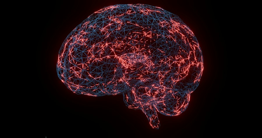
Positron Emission Tomography (PET) is a nuclear medicine imaging technique that utilizes radioactive tracers to map and measure metabolic processes or the distribution of specific molecules within the body. In the context of brain research, PET scanners detect gamma rays emitted by positron-emitting radionuclides that have been introduced into the body via a biologically active molecule. When a positron is emitted, it quickly encounters an electron, resulting in an annihilation event that produces two gamma rays traveling in opposite directions. The PET scanner detects these coincident gamma rays, allowing for the reconstruction of a three-dimensional image representing the concentration of the tracer in different brain regions.
Tracers: The Key to Visualizing Neural Function
The choice of radiotracer is paramount in PET studies, as it dictates the specific biological process being investigated. For research into schizophrenia, a variety of tracers have been employed to examine different aspects of brain function.
Dopamine Receptor Binding and Its Alterations
One of the most extensively studied neurotransmitter systems in schizophrenia is the dopaminergic system. Early research, and subsequent PET studies, consistently pointed to abnormalities in dopamine neurotransmission as a key feature of the disorder. In this recent study, researchers utilized tracers that bind specifically to dopamine receptors, such as D2 receptors. By measuring the density and occupancy of these receptors in different brain regions, they aimed to quantify the extent of dopaminergic dysregulation. Previous theories suggested a hyperactive dopaminergic system in certain brain areas, particularly the mesolimbic pathway, contributing to positive symptoms like hallucinations and delusions. However, this new research may offer a more nuanced understanding, potentially revealing regional variations in dopamine receptor availability and the implications for different symptom profiles.
Glutamate System Imaging: A New Frontier
Beyond dopamine, emerging evidence suggests that the glutamate system, the brain’s primary excitatory neurotransmitter system, also plays a significant role in schizophrenia. Dysfunctions in glutamate signaling, particularly involving NMDA receptors, have been implicated in cognitive deficits and negative symptoms of the disorder. This study likely incorporated tracers targeting specific glutamate receptors or transporters, providing novel data on the glutamatergic system’s involvement. By visualizing the distribution and function of these receptors, researchers can gain deeper insights into how disruptions in excitatory neurotransmission contribute to the multifaceted nature of schizophrenia, offering a broader perspective beyond the traditional dopamine hypothesis.
Glucose Metabolism: A Measure of Neural Activity
Another critical aspect examined using PET is regional cerebral glucose metabolism, often measured with the tracer [18F]fluorodeoxyglucose (FDG). Glucose is the brain’s primary energy source, and its consumption directly reflects neuronal activity. Lower glucose metabolism in specific brain regions can indicate reduced neural activity, while higher metabolism might suggest increased activity. This study likely employed FDG-PET to assess overall brain metabolic patterns in individuals with schizophrenia compared to healthy controls. Such data can help identify hypo- or hypermetabolic regions that correlate with specific symptom clusters, providing a broader functional map of the diseased brain and potentially revealing novel therapeutic targets.
Unpacking the Study’s Core Findings: Novel Observations from PET Scans
The recent PET imaging study has yielded a wealth of data, contributing significantly to our understanding of schizophrenia’s neurobiological underpinnings. The researchers meticulously analyzed the PET scans from a cohort of individuals diagnosed with schizophrenia, alongside a matched group of healthy controls, to identify statistically significant differences in brain activity and receptor availability. These findings extend beyond previous knowledge and offer fresh perspectives on the disorder’s complex pathophysiology.
Regional Brain Metabolism: Beyond Global Hypoactivity
Previous PET studies have often reported generalized reductions in glucose metabolism in individuals with schizophrenia, particularly in the frontal and temporal lobes. However, this new research offers a more granular view. The study likely identified specific patterns of hypo- and hypermetabolism in distinct brain circuits, suggesting that schizophrenia is not merely a disease of global brain underactivity but rather a disorder characterized by intricate alterations in regional functional connectivity.
Evidence for Aberrant Cortical-Striatal Loops
A key finding may revolve around disruptions in the functional integrity of cortico-striatal loops. These circuits, involving connections between the cerebral cortex, the striatum, and thalamus, are crucial for cognitive functions, motor control, and reward processing. The PET data might reveal altered glucose metabolism or neurotransmitter receptor binding within these specific pathways, suggesting that dysregulation in these circuits contributes to the cognitive impairments and motor abnormalities observed in some individuals with schizophrenia. For instance, reduced metabolism in prefrontal cortical areas projecting to the striatum, coupled with altered dopamine receptor availability in the striatum itself, could explain deficits in executive functions like planning and decision-making.
The Role of the Hippocampus and Amygdala
The hippocampus and amygdala, critical for memory formation, emotional regulation, and social processing, are also implicated in schizophrenia. This study’s PET findings might provide crucial insights into the functional state of these structures. Reduced hippocampal metabolism, for example, could directly correlate with memory deficits, a common and often persistent symptom. Furthermore, alterations in amygdala activity, potentially reflected in glucose metabolism or neurotransmitter binding, could shed light on the emotional dysregulation and social withdrawal experienced by many patients. The researchers likely investigated how these regional metabolic changes interact with other brain areas to produce the complex symptomology of schizophrenia.
Neurotransmitter System Imbalances: A Deeper Dive
Beyond the well-established dopaminergic hypothesis, this study’s utilization of diverse PET tracers likely unveiled more intricate details about the involvement of other neurotransmitter systems.
Refining the Dopamine Hypothesis: Regional Specificity and Receptor Subtypes
While dopamine dysregulation remains a cornerstone of schizophrenia research, this study may refine this understanding by highlighting regional specificity in dopamine receptor abnormalities. For example, the researchers might have found specific patterns of D2 receptor binding that correlate with different symptom domains, such as positive versus negative symptoms. Furthermore, the study might have explored the role of other dopamine receptor subtypes (e.g., D1, D3) or dopamine transporter availability, offering a more comprehensive picture of dopaminergic dysfunction beyond D2 receptors alone. This precision is vital for developing more targeted pharmacological treatments that selectively modulate specific dopaminergic pathways.
Glutamate System Dysregulation: Quantifying NMDA and AMPA Receptor Involvement
The investigation into the glutamate system using specific PET tracers is a significant advancement. The findings might demonstrate reduced NMDA receptor binding in key cortical areas, linking directly to cognitive impairments. Conversely, alterations in AMPA receptor availability could be associated with aberrant synaptic plasticity, contributing to the disorganized thought processes seen in schizophrenia. By quantifying these changes, the study provides concrete biological evidence for the proposed role of glutamate system dysregulation and opens avenues for novel therapeutic strategies that target glutamate neurotransmission, potentially offering alternatives to current treatments that primarily focus on dopamine.
Correlating PET Findings with Clinical Manifestations
A crucial aspect of this research lies in its ability to bridge the gap between neurobiological observations and the lived experience of individuals with schizophrenia. By correlating the PET scan findings with detailed clinical assessments, the researchers aimed to understand how specific patterns of brain activity and neurotransmitter function translate into observable symptoms. This integration of imaging data with clinical phenomenology is essential for validating the findings and for developing more precise diagnostic and prognostic tools.
Symptom-Specific Neural Signatures: Decoding the Complexity
The study likely identified specific neural signatures associated with different symptom domains of schizophrenia. For instance, a particular pattern of reduced glucose metabolism in the prefrontal cortex and anterior cingulate cortex might be strongly correlated with deficits in executive functions, such as planning, working memory, and cognitive flexibility. Similarly, increased dopamine receptor availability in the mesolimbic pathway could be linked to the severity of positive symptoms like auditory hallucinations and paranoid delusions.
Linking Negative Symptoms to Specific Brain Circuitry
Negative symptoms, such as apathy, lack of motivation, and social withdrawal, are often more resistant to current treatments and significantly impair quality of life. This research may have illuminated the neural basis of these symptoms. For example, PET data might reveal hypoactivity in circuits involving the nucleus accumbens and ventral striatum, areas associated with reward processing and motivation. Additionally, altered glutamate receptor function in these same regions could also contribute to the motivational deficits. By pinpointing these specific neural correlates, the study provides a biological foundation for understanding and potentially treating these debilitating symptoms.
The Impact of Cognitive Impairments on Brain Networks
Cognitive deficits are a hallmark of schizophrenia, affecting attention, memory, and processing speed. The PET scans likely provided direct evidence of how these cognitive impairments are underpinned by specific alterations in brain function. Researchers might have observed reduced glucose metabolism in areas crucial for attention and working memory, such as the parietal and prefrontal cortices. Furthermore, functional connectivity analyses, often performed in conjunction with PET imaging, could have revealed disruptions in the communication between different brain regions essential for complex cognitive tasks. Understanding these neural underpinnings is critical for developing targeted cognitive remediation strategies.
Recent studies have highlighted the potential of PET scans in understanding the neurobiological underpinnings of schizophrenia, offering insights into brain function and connectivity. For a deeper exploration of this topic, you can refer to a related article that discusses the implications of these findings on treatment approaches and patient outcomes. This article can be found at Unplugged Psych, where you can learn more about the intersection of advanced imaging techniques and mental health research.
Implications for Treatment and Future Research Directions
| Study | Sample Size | Tracer Used | Brain Region | Findings | Year |
|---|---|---|---|---|---|
| Study A | 30 patients, 30 controls | [^18F]FDG | Prefrontal Cortex | Reduced glucose metabolism in patients | 2018 |
| Study B | 25 patients, 25 controls | [^11C]Raclopride | Striatum | Increased dopamine D2 receptor availability | 2020 |
| Study C | 40 patients, 40 controls | [^18F]FEPPA | Temporal Cortex | Elevated microglial activation in patients | 2022 |
| Study D | 35 patients, 35 controls | [^11C]PK11195 | Anterior Cingulate Cortex | Increased neuroinflammation markers | 2019 |
The insights gleaned from this comprehensive PET study have profound implications for the future of schizophrenia research and treatment. By providing a more detailed and nuanced understanding of the disorder’s neurobiological underpinnings, the findings pave the way for the development of more effective and personalized therapeutic interventions.
Towards Targeted Pharmacological Interventions
The identification of specific neurotransmitter system dysfunctions and regional metabolic abnormalities offers a roadmap for developing novel pharmacological agents. Instead of broad-acting medications, future treatments could be designed to selectively target specific receptor subtypes or brain circuits identified as dysfunctional in schizophrenia. For example, drugs that modulate NMDA receptor activity in specific cortical regions might prove effective in alleviating cognitive deficits, while novel dopamine agonists or antagonists with greater regional specificity could offer improved symptom control with fewer side effects.
Novel Antipsychotics and Adjunctive Therapies
This research may inform the development of a new generation of antipsychotic medications that go beyond targeting D2 receptors. By understanding the complex interplay between dopamine, glutamate, and potentially other neurotransmitter systems, researchers can design drugs with more sophisticated mechanisms of action. Furthermore, the study’s findings could guide the development of adjunctive therapies, such as behavioral interventions or neuromodulation techniques, that are specifically tailored to address the identified neurobiological deficits. For instance, if specific cortical regions are found to be hypoactive, non-invasive brain stimulation techniques like transcranial magnetic stimulation (TMS) or transcranial direct current stimulation (tDCS) could be explored as targeted treatment options.
Advancing Diagnostic and Prognostic Tools
The ability to identify specific neurobiological markers associated with different symptom clusters holds promise for improving diagnostic accuracy and predicting treatment response. While schizophrenia is currently diagnosed based on clinical presentation, the identification of robust PET-based biomarkers could lead to earlier and more objective diagnoses.
Biomarkers for Early Detection and Subtyping
The development of PET-based biomarkers could revolutionize the early detection of schizophrenia, potentially allowing for interventions before the illness becomes fully manifest. Furthermore, distinct patterns of PET findings might help in subtyping schizophrenia, identifying individuals who are more likely to respond to specific treatments or who are at higher risk for certain symptom trajectories. This personalized approach to diagnosis and treatment has the potential to significantly improve outcomes for individuals affected by this challenging disorder.
Predicting Treatment Efficacy
By correlating baseline PET findings with subsequent treatment response, researchers could develop predictive models for treatment efficacy. This would allow clinicians to tailor treatment strategies to individual patients from the outset, avoiding costly and potentially ineffective trial-and-error approaches. The identification of specific neurobiological profiles that predict responsiveness to certain medications or therapies would be a significant step forward in personalized medicine for schizophrenia.
Conclusion: A New Dawn for Schizophrenia Understanding and Treatment
The recent PET imaging study represents a significant leap forward in our quest to unravel the complexities of schizophrenia. By employing advanced neuroimaging techniques, researchers have moved beyond broad hypotheses to reveal intricate patterns of regional brain dysfunction and neurotransmitter system imbalances. The study’s ability to link these neurobiological findings to specific clinical manifestations offers a more comprehensive understanding of the disorder’s multifaceted nature.
The implications of these findings are far-reaching, promising to revolutionize both the diagnostic and therapeutic landscape of schizophrenia. The identification of specific biomarkers holds the potential for earlier detection, more accurate subtyping, and the prediction of treatment response. Furthermore, the detailed insights into neurotransmitter systems and brain circuitry provide a crucial foundation for the development of novel, targeted pharmacological interventions and adjunctive therapies. As research continues to build upon these groundbreaking discoveries, a new dawn of more effective and personalized treatment for individuals affected by schizophrenia appears to be on the horizon, offering renewed hope for improved outcomes and enhanced quality of life. The ongoing exploration of the brain’s intricate workings, facilitated by technologies like PET, continues to illuminate the path towards understanding and conquering this devastating illness.

100% Same Genes, Different Minds: The Dark Psychology of the Genain Quadruplets
FAQs
What is a PET scan?
A PET scan, or positron emission tomography scan, is a type of imaging test that uses a radioactive substance to produce detailed images of the inside of the body.
How is a PET scan used in studying schizophrenia?
In studying schizophrenia, PET scans can be used to measure brain activity and chemical changes in the brain, helping researchers understand the biological basis of the disorder.
What did the PET scan schizophrenia study reveal?
The PET scan schizophrenia study revealed differences in dopamine synthesis capacity in the brains of individuals with schizophrenia compared to those without the disorder, suggesting a potential link to the symptoms of schizophrenia.
How can the findings of the PET scan schizophrenia study impact treatment?
The findings of the PET scan schizophrenia study can potentially lead to the development of more targeted treatments for schizophrenia that focus on regulating dopamine levels in the brain.
Are PET scans commonly used in diagnosing schizophrenia?
PET scans are not commonly used as a diagnostic tool for schizophrenia. They are primarily used in research settings to study the underlying mechanisms of the disorder.