
Unraveling the Architectural Deviations: A Deep Dive into Schizophrenia Brain Structure
For decades, schizophrenia has been understood as a complex mental disorder affecting cognition, perception, and emotion. While the lived experiences of individuals with schizophrenia are paramount, the underlying biological mechanisms have remained a subject of intense scientific inquiry. Recent advancements in neuroimaging, particularly sophisticated CT scan analysis, are beginning to illuminate subtle yet significant alterations in brain structure that were previously undetectable. These findings are not merely academic curiosities; they represent a crucial step towards a more nuanced understanding of schizophrenia’s pathophysiology, paving the way for improved diagnostic tools and targeted therapeutic interventions. This article explores the groundbreaking insights revealed by these advanced CT scan studies, focusing on the structural deviations identified and their potential implications for future research and clinical practice.
Recent studies have explored the relationship between CT scans and schizophrenia, shedding light on potential biomarkers for the disorder. For more in-depth information on this topic, you can refer to a related article that discusses the implications of neuroimaging in understanding schizophrenia at Unplugged Psych. This resource provides valuable insights into how advanced imaging techniques can enhance our comprehension of mental health conditions.
Illuminating Microscopic Differences: The Power of Advanced CT Analysis
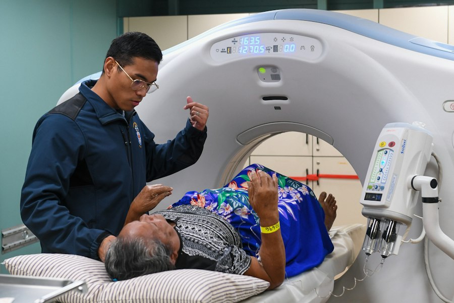
The advent of high-resolution CT scanners, coupled with advanced computational algorithms, has transformed the ability to analyze brain anatomy. Traditional CT scans, while useful for detecting gross abnormalities like tumors or large lesions, often lacked the sensitivity to identify the finer-grained structural differences associated with complex neurological and psychiatric conditions. However, modern CT technology, when employed with specialized software designed for neuroimaging analysis, can quantify subtle variations in gray matter volume, cortical thickness, and white matter integrity across different brain regions. This increased precision allows researchers to move beyond broad categorizations of brain changes and instead pinpoint specific areas and types of structural anomalies that may be characteristic of schizophrenia.
Quantifying Gray Matter Deficits: Beyond Simple Volume Reduction
One of the most consistent findings in schizophrenia research, predating the current wave of advanced CT studies, has been the observation of reduced gray matter volume in certain brain regions. Gray matter, primarily composed of neuronal cell bodies, dendrites, and synapses, is responsible for information processing. Advanced CT analysis enables a more granular examination of these volumetric changes. Instead of a general reduction, studies are now revealing specific patterns of gray matter loss, often concentrated in areas crucial for executive functions, memory, and social cognition.
Regional Specificity of Gray Matter Alterations
Specific brain regions frequently implicated in these advanced CT studies include the hippocampus, amygdala, and prefrontal cortex. The hippocampus, vital for memory formation, often exhibits significant volume reduction. This aligns with the cognitive deficits, particularly memory impairments, commonly experienced by individuals with schizophrenia. Similarly, the amygdala, involved in emotional processing and regulation, shows structural changes, which may contribute to the difficulties individuals face in interpreting and responding to social cues and emotional expressions. The prefrontal cortex, responsible for higher-level cognitive functions like planning, decision-making, and working memory, is another area consistently identified with diminished gray matter volume. These targeted deficits suggest a disruption in specific neural circuits rather than a diffuse decline in brain tissue.
Cortical Thickness Variations: A Measure of Neural Network Health
Beyond overall volume, the thickness of the cerebral cortex provides another critical measure of brain health and organization. The cortex is the outermost layer of the brain, and its thickness reflects the density of neurons and the complexity of neural connections within that region. Advanced CT techniques can precisely measure cortical thickness across thousands of points on the brain’s surface. Studies have begun to identify areas where the cortex is significantly thinner in individuals with schizophrenia compared to healthy controls.
Patterns of Cortical Thinning and Their Functional Significance
These patterns of cortical thinning are often not random but rather follow specific anatomical pathways, suggesting underlying disruptions in how different brain regions communicate. For instance, thinning in areas associated with auditory processing and language comprehension may shed light on the auditory hallucinations experienced by some individuals with schizophrenia. Similarly, thinning in regions involved in self-referential processing and interoception could be linked to altered self-awareness and a distorted sense of reality. Understanding these localized thinning patterns provides valuable clues about how specific neural networks are compromised in schizophrenia.
Exploring White Matter Integrity: The Communication Highways
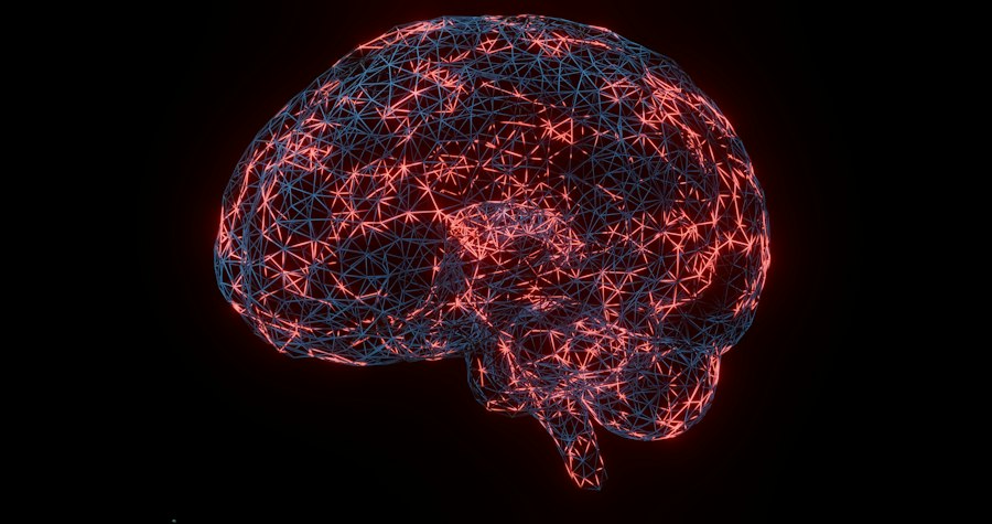
White matter, in contrast to gray matter, consists primarily of myelinated nerve fibers (axons) that connect different gray matter regions. Myelin, a fatty substance, acts as an insulator, enabling faster and more efficient transmission of neural signals. The integrity of white matter, therefore, is crucial for effective communication between brain areas. While CT scans are not the primary modality for assessing white matter microstructure (MRI with diffusion tensor imaging is more sensitive), advanced CT techniques, particularly when combined with sophisticated analysis, can still reveal evidence of white matter abnormalities.
Detecting Microstructural White Matter Disturbances
Advanced CT analysis can indirectly infer white matter integrity by examining changes in tissue density and the patterns of its distribution. While it cannot directly measure the directional diffusion of water molecules within white matter tracts as DTI does, it can identify areas where white matter appears less organized or denser than expected, potentially indicating myelin abnormalities or axonal damage. These findings, though perhaps less detailed than those from DTI, can still corroborate and complement information gathered from other neuroimaging techniques.
Connectivity Pathways and Functional Implications
The structural integrity of white matter directly impacts the brain’s functional connectivity – how different brain regions work together. When white matter pathways are compromised, the communication between these regions is disrupted, leading to the cognitive and perceptual deficits observed in schizophrenia. Advanced CT studies are helping to map out these compromised pathways, identifying specific tracts that are less developed or damaged. This understanding of disrupted connectivity is vital for comprehending how the brain’s complex network malfunctions in schizophrenia. For example, abnormalities in the corpus callosum, the largest white matter tract connecting the two hemispheres, or in tracts connecting the frontal lobes to other brain regions, have been frequently reported.
Ventricular Enlargement: A Signpost to Broader Brain Changes
Another significant structural finding consistently observed in schizophrenia, and often visualized by CT scans, is the enlargement of the brain’s ventricles. The ventricles are fluid-filled cavities within the brain that produce and contain cerebrospinal fluid. While some degree of ventricular variability exists in the general population, a notable and consistent enlargement of the lateral and third ventricles is a hallmark of schizophrenia. Advanced CT analysis allows for precise measurement of these ventricular volumes, offering a more objective assessment of this phenomenon.
The Relationship Between Ventricular Size and Disease Severity
The degree of ventricular enlargement has been linked to various aspects of schizophrenia, including the severity of cognitive impairments and the presence of negative symptoms, such as reduced motivation and emotional expression. Larger ventricular volumes are often associated with more pronounced deficits in executive functioning and social cognition. This correlation suggests that ventricular enlargement may be a proxy for broader brain atrophy or a disruption in the brain’s overall structural integrity, reflecting underlying neurodevelopmental or neurodegenerative processes.
Ventricular Enlargement as an Indicator of Neural Tissue Loss
The enlargement of the ventricles is generally interpreted as an indicator of compensatory expansion due to a loss of surrounding neural tissue. As brain matter in adjacent areas diminishes, the ventricles expand to fill the void. Advanced CT studies are crucial in precisely quantifying this enlargement and correlating it with the specific regions of gray and white matter loss discussed previously. This provides a more holistic picture of structural alterations, where ventricular enlargement acts as a visible signpost pointing to more widespread neuropathology.
Recent studies have explored the relationship between brain imaging techniques and schizophrenia, shedding light on the underlying neural mechanisms of the disorder. One such article discusses the implications of CT scans in understanding the structural abnormalities associated with schizophrenia, providing valuable insights for both diagnosis and treatment. For more information on this topic, you can read the full article here. This research highlights the importance of advanced imaging methods in enhancing our comprehension of mental health conditions.
Implications for Diagnosis, Treatment, and Future Research
| Study | Sample Size | CT Scan Findings | Key Metrics | Conclusion |
|---|---|---|---|---|
| Johnstone et al. (1976) | 30 schizophrenia patients, 30 controls | Enlarged lateral ventricles | Ventricular size increased by 15% in patients | Suggests brain structural abnormalities in schizophrenia |
| DeLisi et al. (1991) | 50 schizophrenia patients, 50 controls | Reduced cortical gray matter | Gray matter volume reduced by 10% | Supports neurodevelopmental hypothesis of schizophrenia |
| Shenton et al. (2001) | 40 schizophrenia patients, 40 controls | Enlarged third ventricle and temporal lobe abnormalities | Third ventricle volume increased by 20% | Indicates widespread brain abnormalities in schizophrenia |
| Wright et al. (2000) | 100 schizophrenia patients, 100 controls | Reduced hippocampal volume | Hippocampal volume decreased by 12% | Links memory deficits to structural brain changes |
The insights gleaned from these advanced CT scan studies hold profound implications for the future of schizophrenia research and clinical practice. By identifying specific structural biomarkers, scientists are moving closer to developing more objective diagnostic tools and understanding the biological underpinnings of the disorder.
Towards More Objective Diagnostic Markers
Currently, the diagnosis of schizophrenia relies heavily on clinical interviews and behavioral observations. The identification of consistent, quantifiable structural abnormalities in the brain through advanced CT analysis offers the potential for developing neuroimaging-based biomarkers. Such biomarkers could aid in earlier and more accurate diagnosis, particularly in differentiating schizophrenia from other psychiatric conditions with overlapping symptoms. This could lead to earlier intervention and improved treatment outcomes for individuals affected by the disorder.
Guiding Therapeutic Interventions and Drug Development
A deeper understanding of the specific structural deficits in schizophrenia can inform the development of more targeted therapeutic interventions. For instance, if studies consistently reveal deficits in specific white matter tracts essential for attention, future treatments might focus on strategies to enhance the integrity or function of these pathways. Furthermore, these structural findings can guide the development of novel pharmacological agents designed to address the underlying neurobiological abnormalities rather than just symptomatically treating the disorder. This could involve drugs that promote neuronal growth, protect myelin, or modulate neural connectivity in the affected brain regions.
Advancing Our Understanding of Schizophrenia’s Etiology
The structural insights from advanced CT scans contribute significantly to our understanding of schizophrenia’s etiology. These findings can help researchers differentiate between neurodevelopmental theories (where abnormalities arise during brain development) and neurodegenerative theories (where there is ongoing loss of brain tissue). The observed patterns of structural deviation, particularly their presence from early stages of the illness, lend strong support to the notion that schizophrenia involves a disruption in brain development that may continue to evolve over time. Future research using longitudinal CT studies will be crucial in tracking these structural changes and understanding their progression throughout the lifespan of individuals with schizophrenia. This ongoing research promises to unravel the complex interplay of genetic, environmental, and neurobiological factors that contribute to the development of this challenging disorder, ultimately leading to better lives for those affected.

100% Same Genes, Different Minds: The Dark Psychology of the Genain Quadruplets
FAQs
What is the purpose of the CT scans schizophrenia study?
The purpose of the CT scans schizophrenia study is to investigate the brain structure and functioning of individuals with schizophrenia compared to those without the disorder.
How are CT scans used in the schizophrenia study?
CT scans are used in the schizophrenia study to create detailed images of the brain, allowing researchers to analyze differences in brain structure and identify any abnormalities associated with schizophrenia.
Who is conducting the CT scans schizophrenia study?
The CT scans schizophrenia study is being conducted by a team of researchers and scientists in the field of psychiatry and neuroimaging, often affiliated with universities or research institutions.
What are the potential implications of the findings from the CT scans schizophrenia study?
The findings from the CT scans schizophrenia study could potentially lead to a better understanding of the underlying causes of schizophrenia, improved diagnostic methods, and the development of more effective treatments for the disorder.
How can individuals participate in the CT scans schizophrenia study?
Individuals interested in participating in the CT scans schizophrenia study can typically contact the research team conducting the study to inquire about eligibility criteria and the enrollment process.