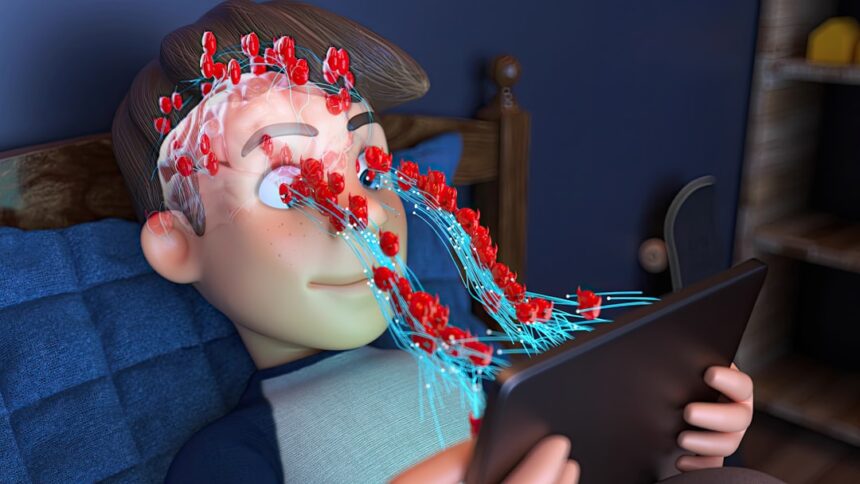
You stand on the precipice of understanding, facing a complex interplay of neural networks, chemical messengers, and behavioral responses. Anxiety, far from a simple emotion, is an elaborate symphony conducted within the confines of your brain. To truly grasp its mechanisms, you must delve into the intricate architecture that underpins your every anxious thought and physiological reaction. This exploration will illuminate not just the origins of your unease, but also potential avenues for navigating its persistent currents.
Imagine your brain as a highly sophisticated security system. The amygdala, a small, almond-shaped structure nestled deep within your temporal lobe, functions as the central alarm bell. Its primary role is to detect threats, both real and perceived, and to initiate a rapid, often reflexive, response. This is not a conscious decision; rather, it’s an ancient, evolutionary mechanism designed for survival.
The Amygdala’s Vigilance: Threat Detection
Your amygdala is constantly scanning your environment, analyzing sensory input for signs of danger. It acts as a lightning-fast filter, prioritizing stimuli that could signify a threat. For example, if you hear a sudden, loud bang, your amygdala immediately flags it as potentially dangerous, even before your conscious mind has fully processed the sound. This pre-conscious processing is a critical component of its efficiency. It doesn’t waste time on careful deliberation when immediate action might be required.
The Role of Sensory Input: A Direct Pathway
Sensory information from your eyes, ears, nose, and touch receptors travels along two distinct pathways to the amygdala. The “low road” is a fast, direct route, bypassing the cortical areas responsible for conscious thought and detailed analysis. This allows for an almost instantaneous threat response. Imagine a flickering shadow in your peripheral vision – your amygdala might trigger a fleeting sense of alarm before you even fully register the object. The “high road,” conversely, sends sensory information to the thalamus, then to your sensory cortex for more detailed processing, and finally to the amygdala. This longer route allows for a more nuanced appraisal of the situation. For instance, if the flickering shadow is identified as a harmless leaf, your amygdala’s initial alarm can be modulated.
Connections to the Hypothalamus and Brainstem: The Fight, Flight, or Freeze Response
When the amygdala detects a threat, it doesn’t operate in isolation. It’s intimately connected to other brain regions that orchestrate your body’s physical and physiological responses. A crucial connection is to the hypothalamus, the command center for your autonomic nervous system. The hypothalamus, in turn, activates the sympathetic nervous system, triggering the classic “fight, flight, or freeze” response. You may feel your heart race, your breathing quicken, and your muscles tense – these are all direct consequences of this intricate neural cascade. Simultaneously, connections to the brainstem can lead to involuntary responses like increased perspiration or dilated pupils. You are experiencing your brain’s ancient survival software taking over.
Recent studies in the neuroscience of anxiety have shed light on the intricate mechanisms underlying this condition, revealing how brain structures like the amygdala and prefrontal cortex interact to regulate fear responses. For a deeper understanding of these findings and their implications for treatment, you can explore the article available at Unplugged Psych, which discusses the latest research and therapeutic approaches in managing anxiety disorders.
The Prefrontal Cortex: The Brain’s Executive Control Center
While the amygdala is designed for rapid threat detection, you also possess a sophisticated executive control center: the prefrontal cortex (PFC). Located at the front of your brain, the PFC is responsible for higher-order cognitive functions such as planning, decision-making, working memory, and, crucially, emotional regulation. It acts as the thoughtful supervisor to the amygdala’s more impulsive alarm.
Working Memory and Contextualization: Assessing the Threat
The PFC plays a vital role in evaluating the true nature of a perceived threat. It accesses your working memory, comparing the current situation with past experiences and knowledge. For example, if your amygdala initially signals alarm at the sound of a fire truck siren, your PFC quickly accesses the information that sirens indicate emergency services, not a direct threat to you personally. This contextualization allows you to modulate your emotional response. Without the PFC, every unexpected sound could trigger a full-blown panic.
Emotional Regulation: Dampening the Alarm
One of the most critical functions of the PFC in the context of anxiety is its ability to regulate emotional responses. It exerts inhibitory control over the amygdala, acting as a brake on its threat-signaling capabilities. When your PFC is fully engaged, you can rationally assess a situation, distinguish between real danger and perceived danger, and consciously choose to calm yourself. This top-down regulation is a hallmark of emotional resilience. However, when the PFC’s influence is weakened, for example, due to chronic stress or certain psychiatric conditions, the amygdala can operate unchecked, leading to heightened anxiety.
The Dialogue Between PFC and Amygdala: A Two-Way Street
The relationship between the PFC and the amygdala is not a one-way street, where the PFC simply dictates terms. It’s a dynamic, interconnected dialogue. While the PFC can inhibit amygdala activity, the amygdala can also influence the PFC. Under extreme stress or chronic anxiety, the amygdala’s constant alarm signals can actually impair the functioning of the PFC, making it harder for you to think clearly, make decisions, and regulate your emotions. This can create a vicious cycle, where anxiety feeds cognitive impairment, which in turn exacerbates anxiety. You might feel “stuck” in a loop of worry, struggling to break free.
Neurotransmitters: The Chemical Messengers of Anxiety
The intricate dance of neural networks in your brain is orchestrated by an array of chemical messengers known as neurotransmitters. These chemicals transit impulses across synapses, the tiny gaps between neurons, influencing everything from your mood and motivation to your perception of fear. Imbalances in these crucial chemicals are often implicated in the experience of anxiety.
Serotonin: The Mood Regulator
Serotonin is often referred to as the “feel-good” neurotransmitter, playing a significant role in regulating mood, sleep, appetite, and social behavior. Low levels or dysfunctional serotonin pathways are frequently associated with anxiety and depression. When serotonin levels are optimized, you experience a greater sense of calm and well-being. Many antidepressant medications, known as selective serotonin reuptake inhibitors (SSRIs), work by increasing the availability of serotonin in the synaptic cleft, thereby enhancing its signaling and often alleviating anxiety symptoms. Imagine serotonin as the lubricant for your emotional gears; when it’s flowing smoothly, your emotional machinery operates efficiently.
GABA: The Brain’s Natural Tranquilizer
Gamma-aminobutyric acid, or GABA, is the primary inhibitory neurotransmitter in your brain. Its primary function is to calm neural activity, counteracting the excitatory effects of other neurotransmitters. Think of GABA as your brain’s natural tranquilizer. When GABA binds to its receptors, it reduces the excitability of neurons, leading to a state of relaxation and reduced anxiety. Individuals with anxiety disorders often exhibit decreased GABA activity or GABA receptor dysfunction, suggesting a compromised capacity for natural calming. Benzodiazepines, a class of anti-anxiety medications, enhance the effects of GABA, producing a rapid calming effect.
Norepinephrine: The Arousal Amplifier
Norepinephrine, also known as noradrenaline, is both a neurotransmitter and a hormone. It plays a key role in your “fight-or-flight” response, increasing heart rate, vigilance, and glucose release. While essential for alertness and focus, an overactive norepinephrine system can contribute significantly to anxiety symptoms. You might experience a racing heart, sweaty palms, and a sense of hyper-vigilance, feeling constantly on edge. This neurotransmitter essentially puts your body and mind on high alert, even in the absence of a genuine threat. It’s like turning up the volume on your internal alarm system to an unbearable degree.
Dopamine: Reward and Motivation, But Also Anxiety
Dopamine is primarily associated with the brain’s reward system, motivation, and pleasure. However, its role in anxiety is more complex and nuanced. While it can contribute to a sense of well-being, dysregulation of dopamine pathways can also be implicated in anxiety. For instance, imbalances in dopamine can contribute to worry, fear, and even panic attacks in some individuals. Its dynamic interplay with other neurotransmitters means that while it brings pleasure, an excess or deficiency in certain pathways can throw your emotional equilibrium off balance.
The Role of Stress Hormones: Cortisol and Adrenaline
Beyond neurotransmitters, your brain also orchestrates the release of powerful hormones that significantly impact your anxiety levels. The hypothalamic-pituitary-adrenal (HPA) axis, often referred to as the stress axis, is a complex neuroendocrine system that plays a central role in your body’s response to stress.
The Hypothalamic-Pituitary-Adrenal (HPA) Axis: Stress Orchestrator
When you perceive a stressor, your hypothalamus releases corticotropin-releasing hormone (CRH). This signals your pituitary gland to release adrenocorticotropic hormone (ACTH), which then travels through your bloodstream to your adrenal glands. The adrenal glands, located atop your kidneys, then release a surge of stress hormones: cortisol and adrenaline. This cascade is designed to prepare your body for immediate action.
Cortisol: The Sustained Stress Response
Cortisol is the primary stress hormone and is often referred to as the body’s natural alarm clock. It fuels your body for action by increasing blood sugar, suppressing non-essential bodily functions, and enhancing memory for emotionally significant events. While crucial for acute stress, chronically elevated cortisol levels, often seen in prolonged anxiety, can have detrimental effects. These include impaired cognitive function (especially in the hippocampus, a region crucial for memory), weakened immune system, and increased risk for various physical and mental health issues. Imagine cortisol as a sustained energy boost, but one that, if left unchecked, can burn out your system.
Adrenaline (Epinephrine): The Immediate Rush
Adrenaline, or epinephrine, is another key stress hormone released during the “fight-or-flight” response. It provides a rapid surge of energy, constricts blood vessels in some areas while dilating them in others, increases heart rate and blood pressure, and prepares your muscles for immediate action. The sudden rush of adrenaline can feel like a jolt, causing symptoms such as palpitations, shortness of breath, and a feeling of impending doom. While beneficial in a genuine emergency, recurrent adrenaline surges in the absence of real danger contribute significantly to the distressing physical symptoms of anxiety and panic attacks. This is the immediate, explosive energy burst that your body deploys.
Recent research in the neuroscience of anxiety has shed light on the complex interplay between brain structures and emotional regulation. For those interested in exploring this topic further, a related article discusses how various neural pathways contribute to anxiety disorders and potential therapeutic approaches. You can read more about these findings in the insightful piece available at Unplugged Psych, which delves into the mechanisms behind anxiety and offers a comprehensive overview of current treatment options.
Neuroplasticity and Anxiety: The Malleable Brain
| Metric | Description | Typical Findings in Anxiety | Measurement Method |
|---|---|---|---|
| Amygdala Activation | Level of activity in the amygdala, a brain region involved in fear processing | Increased activation in response to threat-related stimuli | fMRI (functional Magnetic Resonance Imaging) |
| Prefrontal Cortex Regulation | Activity in the prefrontal cortex responsible for top-down control of emotions | Reduced activation or connectivity with amygdala | fMRI, EEG (Electroencephalography) |
| Heart Rate Variability (HRV) | Variability in time intervals between heartbeats, indicating autonomic nervous system balance | Lower HRV indicating higher sympathetic (stress) activity | ECG (Electrocardiogram) |
| Cortisol Levels | Concentration of cortisol hormone related to stress response | Elevated baseline or reactive cortisol levels | Saliva or blood assays |
| GABA Concentration | Levels of gamma-aminobutyric acid, the main inhibitory neurotransmitter | Reduced GABA levels in certain brain regions | MRS (Magnetic Resonance Spectroscopy) |
| Skin Conductance Response (SCR) | Changes in skin electrical conductance due to sweat gland activity | Increased SCR indicating heightened arousal | Electrodermal activity sensors |
Your brain is not a static organ; it is constantly changing and adapting in response to your experiences. This remarkable ability, known as neuroplasticity, means that neural pathways can be strengthened or weakened, new connections can be formed, and existing ones can be pruned. This inherent malleability has profound implications for understanding and addressing anxiety.
Strengthening Anxious Pathways: The Learning of Fear
When you repeatedly experience anxious thoughts or situations, the neural pathways associated with fear and anxiety become more established and efficient. It’s like repeatedly traversing a path through a dense forest; with each journey, the path becomes clearer and easier to follow. If you consistently ruminate on worries, for example, your brain literally gets better at worrying. This “learning” of anxiety can lead to a state where your brain automatically defaults to anxious responses, even in situations that are not inherently threatening.
The Impact of Early Life Experiences: Shaping the Brain
Early life experiences, particularly those involving trauma or chronic stress, can significantly shape the development of your brain’s anxiety circuits. Adverse childhood experiences can lead to alterations in brain regions involved in emotional regulation, such as the amygdala and prefrontal cortex, making individuals more vulnerable to anxiety later in life. These experiences can essentially “hardwire” a heightened threat response, making it more challenging to manage anxious feelings as an adult. Your early environment acts as a sculptor, molding the very architecture of your emotional landscape.
Rewiring Anxious Brains: Therapeutic Interventions
The good news is that neuroplasticity also offers a powerful avenue for therapeutic intervention. Your brain’s ability to change means that anxious pathways can be weakened, and new, more adaptive pathways can be strengthened. Therapies like Cognitive Behavioral Therapy (CBT) work by challenging negative thought patterns and maladaptive behaviors, thereby actively rewiring your brain. Mindfulness practices teach you to observe anxious thoughts without engaging with them, gradually reducing their power. Even lifestyle changes, such as exercise and a healthy diet, can foster positive neuroplastic changes, promoting resilience and reducing anxiety. You are not condemned to your existing neural pathways; you have the power to reshape them. This takes conscious effort and consistent practice, much like building a new muscle.
FAQs
What is the neuroscience of anxiety?
The neuroscience of anxiety studies how brain structures, neural circuits, and biochemical processes contribute to the experience and regulation of anxiety. It explores how different brain regions, such as the amygdala and prefrontal cortex, interact to produce anxiety responses.
Which brain areas are primarily involved in anxiety?
Key brain areas involved in anxiety include the amygdala, which processes fear and threat detection; the prefrontal cortex, which regulates emotional responses; the hippocampus, which is involved in memory and context; and the hypothalamus, which controls stress hormone release.
How do neurotransmitters affect anxiety?
Neurotransmitters like gamma-aminobutyric acid (GABA), serotonin, norepinephrine, and dopamine play crucial roles in anxiety. For example, reduced GABA activity can lead to increased anxiety, while serotonin helps regulate mood and anxiety levels.
Can brain imaging techniques help understand anxiety?
Yes, brain imaging methods such as functional magnetic resonance imaging (fMRI) and positron emission tomography (PET) allow researchers to observe brain activity and connectivity patterns associated with anxiety, helping to identify neural mechanisms and potential treatment targets.
How does understanding the neuroscience of anxiety contribute to treatment?
Understanding the neural basis of anxiety helps in developing targeted therapies, including medications that modulate neurotransmitter systems and behavioral interventions like cognitive-behavioral therapy (CBT) that can alter brain function and connectivity to reduce anxiety symptoms.