
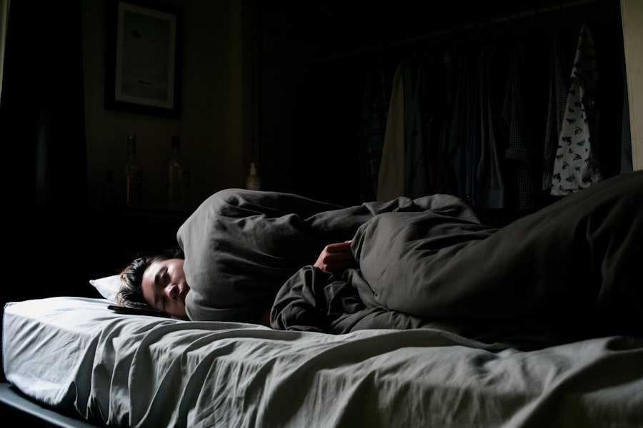
You’ve probably experienced it, or at least heard tales of it: the chilling moment you wake up, fully conscious, yet utterly unable to move. Your eyes dart around the room, your mind screams, but your body remains a leaden weight. This phenomenon, known as sleep paralysis, is a disquieting intruder into the liminal space between wakefulness and sleep. To truly understand this experience, you must delve into the intricate workings of your brain, observing how complex neurological processes can sometimes misfire, creating a temporary state of profound helplessness.
To grasp sleep paralysis, you first need to comprehend the two main stages of sleep: Non-Rapid Eye Movement (NREM) and Rapid Eye Movement (REM) sleep. Think of these as two distinct cities your brain visits each night, each with its own unique architecture and activity.
Non-Rapid Eye Movement (NREM) Sleep
NREM sleep is the deep, restorative phase. During this period, your brain activity slows considerably. You transition through three sub-stages:
- N1 (Light Sleep): This is your gateway to sleep. Your brain activity begins to decelerate, and you may experience hypnagogic jerks, those sudden twitches that feel like you’re falling.
- N2 (True Sleep): Your body temperature drops, your heart rate slows, and your brain produces “sleep spindles” and “K-complexes,” unique patterns visible on an electroencephalogram (EEG) that help keep you asleep and consolidate memories.
- N3 (Deep Sleep/Slow-Wave Sleep): This is the most profound stage of NREM. Your brain waves become very slow and large (delta waves). This is when tissue repair, growth, and immune system strengthening occur. Awakening from N3 sleep often leaves you feeling disoriented or groggy.
Rapid Eye Movement (REM) Sleep
REM sleep is a stark contrast to NREM. It’s the stage where dreams flourish, and your brain activity remarkably resembles that of wakefulness. However, there’s a critical difference.
- Brain Activity: Your brain is highly active, consuming significant energy. The primary visual cortex, responsible for processing sights, lights up, creating vivid nocturnal landscapes.
- Physiological Changes: Your heart rate and breathing become irregular, similar to when you’re awake and active. Your eyes dart rapidly beneath your eyelids, hence the name.
- Muscle Atonia: Crucially, during REM sleep, your brain actively paralyzes most of your voluntary muscles. This is a protective mechanism, preventing you from physically acting out your dreams. Imagine a brain that allows you to fly off your bed trying to evade a dreamed-of monster – chaos would ensue. This muscle paralysis is central to understanding sleep paralysis.
Sleep paralysis is a fascinating phenomenon that has intrigued both scientists and the general public alike, often linked to the neuroscience of sleep and the brain’s mechanisms during REM sleep. For a deeper understanding of this subject, you can explore a related article that delves into the intricacies of sleep paralysis and its neurological underpinnings. This article provides insights into the experiences of individuals who have encountered sleep paralysis and discusses the potential causes and implications for mental health. To read more, visit this article.
The Mechanism of REM Atonia: A Brain’s Safety Switch
The temporary paralysis you experience during sleep paralysis is not a malfunction in itself, but rather an exaggeration or mis-timing of a normal physiological process: REM atonia. Your brain, with its extraordinary complexity, has a sophisticated “safety switch” to keep you from harming yourself while you dream.
Neurotransmitter Systems Involved
The meticulous orchestration of REM atonia involves a complex interplay of neurotransmitters.
- GABA (Gamma-Aminobutyric Acid): This is your brain’s primary inhibitory neurotransmitter. During REM sleep, specific neurons in the brainstem, particularly in the pontine reticular formation, become highly active and release GABA. This increased GABA activity effectively “puts the brakes” on motor neurons in the spinal cord, preventing them from firing.
- Glycine: Alongside GABA, glycine acts as another inhibitory neurotransmitter, amplifying the paralyzing effect on motor neurons. Think of GABA and glycine as two skilled technicians working in tandem to shut down your muscle control.
- Acetylcholine: While GABA and glycine inhibit motor activity, acetylcholine plays a crucial role in activating other aspects of REM sleep, such as dream generation and rapid eye movements. The intricate balance between these excitatory and inhibitory neurotransmitters is key to proper sleep cycling.
The Brainstem’s Role
The brainstem, a primitive but vital part of your brain, acts as the control center for many basic life functions, including sleep-wake cycles. Within the brainstem, specific nuclei are responsible for initiating and maintaining REM atonia. Lesions or disruptions in these critical areas can lead to a condition called REM sleep behavior disorder, where individuals do act out their dreams because the atonia mechanism is compromised. Sleep paralysis, however, represents the other side of this coin – an overzealous or misaligned atonia.
When the Lines Blur: The Pathophysiology of Sleep Paralysis
Sleep paralysis occurs when components of REM sleep intrude into wakefulness. Imagine your brain as a finely tuned orchestra. During sleep paralysis, the “REM atonia” section of the orchestra continues to play its paralyzing tune even after the “wakefulness” section has begun its performance. You are conscious, your sensory perception is active, but your motor commands are muted.
Isolated Sleep Paralysis (ISP)
ISP is the most common form, occurring in individuals without an underlying sleep disorder. It’s often triggered by:
- Irregular Sleep Schedule: Jet lag, shift work, or simply inconsistent bedtimes can disrupt your brain’s natural rhythm.
- Sleep Deprivation: Not getting enough sleep can throw your brain’s delicate balance off kilter.
- Stress and Anxiety: High levels of psychological distress can influence sleep architecture.
- Sleeping Position: Many individuals report experiencing sleep paralysis more frequently when sleeping on their back (supine position).
Recurrent Isolated Sleep Paralysis (RISP)
Some individuals experience ISP frequently, classifying it as RISP. If you find yourself in this situation, it is important to note that while distressing, it is generally considered benign. However, frequent episodes warrant exploring lifestyle adjustments and potentially discussing it with a healthcare professional.
Narcolepsy-Related Sleep Paralysis
Sleep paralysis is also a cardinal symptom of narcolepsy, a chronic neurological disorder characterized by overwhelming daytime sleepiness and other REM sleep-related phenomena. In narcolepsy, the boundaries between wakefulness and sleep are particularly porous. For individuals with narcolepsy, sleep paralysis, along with cataplexy (sudden loss of muscle tone triggered by strong emotions), hypnagogic hallucinations (vivid hallucinations at sleep onset), and hypnopompic hallucinations (vivid hallucinations upon waking), are all manifestations of REM intruding into wakefulness.
The Hallucinatory Landscape: A Mind’s Primal Fear
Beyond the paralysis, many people report terrifying hallucinations during sleep paralysis. These are not dreams in the traditional sense, but rather vivid sensory experiences that occur while you are consciously aware. Think of them as echoes of REM sleep’s dream machinery bleeding into your waking perception.
Hypnagogic and Hypnopompic Hallucinations
These terms distinguish hallucinations occurring at sleep onset (hypnagogic) from those occurring upon waking (hypnopompic). During sleep paralysis, you are primarily experiencing hypnopompic hallucinations.
- Intruder Hallucinations: You might feel a menacing presence in the room, see shadowy figures, or even hear footsteps or whispers. This is arguably the most common and terrifying type, tapping into primal fears of vulnerability.
- Incubus/Succubus Hallucinations: Historically linked to demons or evil spirits, these involve a sense of pressure on the chest, difficulty breathing, and a feeling of being ‘ridden’ or crushed. This sensation is a physiological consequence of the brain misinterpreting the body’s normal REM-atonia induced shallow breathing and suppressed muscle movement as an external force.
- Vestibular-Motor Hallucinations: You may experience sensations of floating, falling, or out-of-body experiences. These are thought to arise from the brain’s internal models of body position and movement becoming disoriented due to the conflicting signals of wakefulness and muscle paralysis.
The Amygdala and Fear Response
During these hallucinatory experiences, your amygdala – the brain’s fear center – is highly activated. Because you are conscious and unable to move, your brain interprets the unusual sensory input as a genuine threat. This triggers a powerful fight-or-flight response, flooding your body with adrenaline and amplifying the terror, even though no actual danger exists externally. This is why sleep paralysis can feel so profoundly terrifying; your brain is convinced you are in imminent peril.
Sleep paralysis, a phenomenon that occurs during the transition between wakefulness and sleep, has intrigued both scientists and the general public alike. Recent research has shed light on the neuroscience behind this unsettling experience, revealing how disruptions in REM sleep can lead to the temporary inability to move or speak. For those interested in exploring this topic further, an insightful article can be found at Unplugged Psychology, which delves into the psychological and physiological aspects of sleep paralysis, offering a comprehensive overview of its causes and effects.
Managing and Preventing Sleep Paralysis
| Metric | Description | Typical Values/Findings | Source/Study |
|---|---|---|---|
| Prevalence | Percentage of general population experiencing sleep paralysis at least once | 8% – 50% | Sharpless & Barber, 2011 |
| Average Duration | Length of a typical sleep paralysis episode | 30 seconds to 2 minutes | Cheyne et al., 1999 |
| REM Sleep Atonia | Muscle paralysis during REM sleep, underlying mechanism of sleep paralysis | Complete skeletal muscle atonia except diaphragm and eye muscles | Hobson et al., 2000 |
| Brain Activity During Episode | Neural correlates observed during sleep paralysis episodes | Increased activity in amygdala and parietal cortex | Jalal et al., 2017 |
| Common Triggers | Factors increasing likelihood of sleep paralysis | Sleep deprivation, irregular sleep schedule, stress | Denis et al., 2018 |
| Emotional Response | Typical feelings reported during episodes | Fear (up to 90%), hallucinations (visual/auditory) | Cheyne, 2005 |
| Genetic Influence | Heritability estimate of sleep paralysis susceptibility | Estimated 28% heritability | Denis et al., 2015 |
While sleep paralysis can be a profoundly disturbing experience, you are not powerless against it. Understanding its neurological underpinnings can empower you to take steps to minimize its occurrence and lessen its impact.
Lifestyle Adjustments
Many cases of sleep paralysis are linked to disrupted sleep patterns. You can significantly reduce your chances of experiencing it by adopting healthy sleep hygiene practices.
- Maintain a Regular Sleep Schedule: Go to bed and wake up at roughly the same time each day, even on weekends. This helps regulate your body’s internal clock, your circadian rhythm.
- Ensure Adequate Sleep: Aim for 7-9 hours of quality sleep per night. Sleep deprivation is a major trigger.
- Create a Conducive Sleep Environment: Your bedroom should be dark, quiet, and cool.
- Limit Stimulants: Avoid caffeine and nicotine, especially in the hours leading up to bedtime.
- Avoid Alcohol: While alcohol may initially make you feel drowsy, it disrupts sleep architecture later in the night, often leading to more fragmented sleep and increased REM rebound.
- Manage Stress: Incorporate relaxation techniques into your daily routine, such as meditation, yoga, or deep breathing exercises. High stress levels can easily throw your sleep cycle off.
- Adjust Sleeping Position: If you frequently experience ISP, try sleeping on your side. For many, sleeping on their back seems to increase the likelihood of an episode.
During an Episode: Coping Strategies
When an episode strikes, it can feel like an eternity. However, you can employ certain techniques to try and break free.
- Focus on Small Movements: Attempt to wiggle a finger or a toe. These smaller muscles are sometimes less affected by the atonia, and initiating movement, no matter how tiny, can sometimes “wake up” the rest of your motor system.
- Try to Make a Sound: If even a whisper is possible, the effort can sometimes help you fully awaken.
- Relax and Wait It Out: Paradoxically, panicking often prolongs the episode. Remind yourself it’s temporary and harmless, a mere trick of the mind. Focus on your breathing and ride the wave until it subsides.
- Don’t Re-enter Sleep Immediately: Once you break free, get up, walk around, or briefly engage in a non-stimulating activity before attempting to go back to sleep. This helps ensure your brain fully exits the REM state.
Seeking Professional Help
If sleep paralysis is recurrent, significantly impacts your quality of life, or is accompanied by excessive daytime sleepiness, it’s crucial to consult a healthcare professional.
- General Practitioner: Your GP can rule out any underlying medical conditions and provide initial advice.
- Sleep Specialist: If necessary, your doctor may refer you to a sleep specialist who can conduct a thorough evaluation, potentially including a sleep study (polysomnography). This can identify or rule out conditions like narcolepsy or other sleep disorders.
- Therapy: Cognitive Behavioral Therapy for Insomnia (CBT-I) or other forms of counseling can be helpful in managing sleep-related anxiety and improving sleep hygiene.
Understanding the neuroscience behind sleep paralysis transforms a terrifying, inexplicable experience into a fascinating, albeit inconvenient, glimpse into the complex machinery of your sleeping brain. You are not alone, you are not going insane, and with knowledge and appropriate strategies, you can minimize the unsettling intrusions of the night.
FAQs
What is sleep paralysis?
Sleep paralysis is a temporary inability to move or speak that occurs when a person is falling asleep or waking up. It happens during the transition between wakefulness and rapid eye movement (REM) sleep, when the brain is active but the body remains immobile.
What causes sleep paralysis from a neuroscience perspective?
Sleep paralysis is caused by a disruption in the normal sleep cycle, particularly during REM sleep. During REM, the brain sends signals to inhibit muscle activity to prevent acting out dreams. If the brain wakes up before this inhibition ends, the person becomes conscious but unable to move, resulting in sleep paralysis.
Are hallucinations common during sleep paralysis?
Yes, hallucinations are common during sleep paralysis. These can include visual, auditory, or tactile sensations and are thought to arise from the brain’s heightened activity in areas related to fear and perception, combined with the inability to move.
Is sleep paralysis harmful to the brain or body?
Sleep paralysis itself is not harmful to the brain or body. It is generally a benign phenomenon, although it can be frightening. However, frequent episodes may be associated with sleep disorders or mental health conditions and should be evaluated by a healthcare professional.
Can sleep paralysis be prevented or treated?
Improving sleep hygiene, reducing stress, and maintaining a regular sleep schedule can help reduce the frequency of sleep paralysis episodes. In some cases, treating underlying sleep disorders or anxiety may be necessary. There is no specific medication for sleep paralysis, but addressing contributing factors is effective.