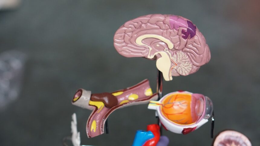
You are about to delve into the intricate world of perineuronal nets (PNNs), a fascinating and increasingly recognized component of the adult brain. These specialized extracellular matrix structures, far from being mere scaffolding, are emerging as critical regulators of neuronal function and plasticity, particularly in the mature nervous system. You will discover how PNNs act as both guardians of stability and facilitators of change, much like a carefully managed botanical garden that maintains its overall structure while allowing for the subtle growth and adaptation of individual plants.
To truly comprehend the role of PNNs, you must first understand their fundamental composition and architectural arrangement around neurons. These structures are not ubiquitous throughout the brain but exhibit a distinct preference for specific neuronal populations, suggesting a highly targeted function.
Compositional Complexity: The Molecular Building Blocks
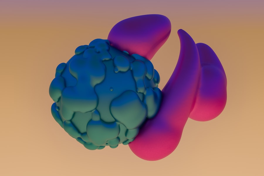
Imagine a sophisticated molecular mesh, woven with precision around certain neurons. This is, in essence, a PNN. You will find that these nets are primarily composed of a complex interplay of several key molecular families, each contributing to their unique properties and functions.
Hyaluronan: The Foundational Polymer
At the heart of the PNN lies hyaluronan (HA), a large, unbranched glycosaminoglycan. Think of HA as the long, flexible backbone of the net, providing its overall structural integrity. Its intrinsic properties, such as its ability to attract and retain water, contribute to the PNN’s viscoelastic nature. You will observe that the length and cross-linking of HA molecules significantly influence the density and permeability of the net.
Chondroitin Sulfate Proteoglycans (CSPGs): The Regulatory Elements
Interspersed within the HA framework are chondroitin sulfate proteoglycans (CSPGs), a diverse family of molecules that are arguably the most functionally dynamic components of PNNs. These proteoglycans, such as aggrecan, brevican, neurocan, and versican, possess core proteins adorned with numerous chondroitin sulfate (CS) chains. You will come to appreciate that the sulfation patterns on these CS chains are not random but highly specific, acting like molecular barcodes that can influence interactions with other molecules, including growth factors, receptors, and adhesion molecules. Consider these CS chains as the nuanced control knobs of the PNN, fine-tuning its interactions and regulatory capacity.
Link Proteins: Stabilizers and Connectors
Just as reinforced concrete relies on steel rebar for strength, PNNs utilize link proteins to stabilize the association between HA and CSPGs. Specifically, you will encounter the brain-specific link proteins (Bral1 and Bral2) which act as crucial connectors, mediating the tight binding of CSPGs to the HA backbone. This linkage is vital for maintaining the structural integrity of the net, preventing its disaggregation and ensuring its long-term stability around the neuron.
Tenascin-R: A Multifunctional Adhesion Molecule
Another significant component you will observe in PNNs is Tenascin-R. This extracellular matrix glycoprotein plays a role in cell adhesion, migration, and neurite outgrowth. Within the PNN, Tenascin-R is believed to contribute to the overall rigidity and stability of the net, while also potentially modulating neuron-glia interactions. Its presence underscores the multifaceted nature of PNNs, extending beyond mere structural support to a more active role in cellular communication.
Targeted Distribution: A Selective Embrace
PNNs do not indiscriminately ensheath all neurons. You will find that their distribution is highly selective, predominantly surrounding inhibitory interneurons, particularly parvalbumin-positive (PV+) interneurons. This selectivity is a critical clue to their function.
Ensheathment of PV+ Interneurons: A Pervasive Pattern
The strong association of PNNs with PV+ interneurons in various brain regions, including the neocortex, hippocampus, and cerebellum, is a recurring theme in your exploration. These inhibitory interneurons are crucial for regulating neuronal circuit activity, establishing precise timing, and maintaining the excitation-inhibition balance. You will come to see the PNN as a kind of sophisticated “privilege badge” for these essential inhibitory neurons, enhancing their functional robustness.
Functional Implications of Selective Ensheathment
The preferential ensheathment of PV+ interneurons suggests that PNNs play a specific role in modulating the function of these crucial inhibitory cells. By influencing the synaptic environment around PV+ interneurons, PNNs can directly impact the strength and plasticity of inhibitory synapses, thereby regulating the overall excitability of local circuits. You can think of it as the PNN acting as a custom-tailored suit that empowers these inhibitory gatekeepers to perform their duties with greater precision and resilience.
Recent research has highlighted the significance of perineuronal nets in regulating adult brain plasticity, suggesting that these structures play a crucial role in the maturation and stabilization of synapses. An insightful article that delves deeper into this topic can be found at Unplugged Psychology, where the intricate relationship between perineuronal nets and neural adaptability is explored, shedding light on potential implications for cognitive health and recovery from neurological disorders.
Gatekeepers of Critical Periods: Shaping Early Brain Development
While the focus here is on the adult brain, it’s essential to briefly acknowledge the profound role PNNs play during brain development, particularly in the closure of critical periods. This developmental context provides a crucial foundation for understanding their continued influence in adulthood.
Critical Period Maturation: A Temporal Boundary
Critical periods are developmental windows during which the brain exhibits heightened sensitivity to environmental input, allowing for rapid and precise circuit refinement. You will observe that the emergence of PNNs often coincides with the closure of these critical periods, acting as a “brake” on excessive plasticity.
Visual Cortex Plasticity: A Classic Example
The visual cortex offers a compelling illustration. In early development, ocular dominance plasticity, where the visual cortex rapidly adapts to early visual experience, is pronounced. As PNNs mature around inhibitory interneurons during later development, you will find that this plasticity becomes significantly restricted. This suggests that PNNs contribute to the stabilization of established circuits, preventing them from being excessively perturbed by new sensory input. Imagine the PNN as a carefully erected fence that designates the boundaries of a developing neural pathway, ensuring its architectural integrity once its basic structure is laid down.
Molecular Mechanisms of Critical Period Closure
The molecular machinery of PNNs, particularly the chondroitin sulfate chains, is implicated in this critical period closure. You will discover that enzymatic degradation of these CS chains, for example with chondroitinase ABC, can reopen critical period-like plasticity in the adult brain. This highlights the dynamic and potentially reversible nature of PNN-mediated circuit stabilization.
Regulators of Adult Synaptic Plasticity: Maintaining the Balance
In the adult brain, PNNs transitions from primarily closing critical periods to dynamically regulating synaptic plasticity. They are not rigid structures that completely abolish change but rather act as sophisticated modulators, permitting adaptive plasticity while preventing uncontrolled reorganization.
Impeding Excessive Plasticity: A Stabilizing Force
You will appreciate that PNNs contribute to the overall stability of neural circuits by regulating synaptic plasticity. They act as guardians of established synaptic connections, preventing their unwarranted degradation or excessive modification.
Regulating Long-Term Potentiation (LTP) and Depression (LTD)
PNNs have been shown to modulate both long-term potentiation (LTP) and long-term depression (LTD), the cellular mechanisms underlying learning and memory. You will find evidence that PNNs can constrain the induction and magnitude of LTP, particularly at glutamatergic synapses onto PV+ interneurons. Conversely, they can also influence LTD, suggesting a nuanced role in maintaining the balance between synaptic strengthening and weakening. Think of PNNs as traffic controllers for synaptic changes, ensuring that the flow of information is optimized without leading to congestion or collapse.
Influence on Excitatory-Inhibitory Balance
The targeted distribution of PNNs around inhibitory interneurons underscores their crucial role in maintaining the excitation-inhibition (E/I) balance within neural circuits. By fine-tuning the efficacy and plasticity of inhibitory synapses, PNNs indirectly influence the excitability of principal neurons and the overall network activity. You can envision PNNs as sophisticated thermostats, constantly adjusting the E/I ratio to maintain optimal brain function.
Facilitating Specific Forms of Plasticity: Adapting with Precision
While often perceived as restrictive elements, you will find compelling evidence that PNNs also actively facilitate certain forms of adaptive plasticity, illustrating their dual role as both gatekeepers and enablers.
Metaplasticity and Learning
PNNs are increasingly implicated in metaplasticity, which refers to the plasticity of synaptic plasticity. They can influence the threshold for inducing LTP or LTD, effectively “priming” synapses for future changes based on prior activity. This suggests that instead of simply blocking plasticity, PNNs contribute to a more nuanced and context-dependent form of learning. Consider the PNN as a conductor, guiding the orchestra of synaptic changes to produce a harmonious and adaptive performance.
Role in Motor Learning and Skill Acquisition
You will also encounter studies demonstrating a link between PNN integrity and motor learning. For instance, the degradation of PNNs in specific motor regions can facilitate the acquisition of new motor skills, particularly those requiring fine motor control. This suggests that while PNNs stabilize established motor programs, their temporary removal can “unlock” a window of increased plasticity conducive to skill acquisition. This is akin to temporarily removing a meticulously maintained trellis to allow a climbing plant to find a new, more optimal path.
PNNs in Neurological Disorders: A Vulnerable Guardian

The vital roles of PNNs in maintaining brain function and plasticity make them highly relevant to the etiology and progression of various neurological and psychiatric disorders. You will observe their consistent involvement across a spectrum of conditions.
Schizophrenia and Other Psychiatric Disorders
Dysregulation of PNNs has been consistently implicated in schizophrenia. You will encounter studies reporting altered PNN density and compositional changes in the brains of individuals with schizophrenia, particularly in regions associated with cognitive function and executive control.
PNN Deficiencies and Inhibitory Dysfunction
Reduced PNN integrity around PV+ interneurons is often associated with impaired inhibitory neurotransmission, leading to an imbalance of excitation and inhibition, a hallmark of schizophrenia. You can imagine that if the “privilege badge” of these inhibitory neurons is compromised, their ability to regulate neural activity is diminished, contributing to the chaotic neural signaling observed in the disorder.
Therapeutic Potential: Targeting PNNs
The association between PNN dysfunction and schizophrenia suggests potential therapeutic avenues. Modulating PNN components, for example, by promoting their formation or altering their composition, could represent novel strategies for restoring E/I balance and improving cognitive function in affected individuals.
Alzheimer’s Disease and Neurodegeneration
In the context of neurodegenerative diseases like Alzheimer’s, PNNs also exhibit significant alterations. You will find evidence of PNN degradation and a reduction in their numbers in affected brain regions.
Loss of Neuroprotection
PNNs are believed to offer a degree of neuroprotection, shielding neurons from various stressors, including oxidative stress and excitotoxicity. Their degradation in Alzheimer’s disease could therefore exacerbate neuronal vulnerability and contribute to the progression of neurodegeneration. Think of the PNN as a protective suit that, when damaged, leaves the neuron exposed to a hostile environment.
Modulating Amyloid-Beta Pathology
Some research suggests that PNNs can interact with amyloid-beta plaques, the pathological hallmarks of Alzheimer’s disease. The exact nature of this interaction is still under investigation, but it highlights a potential role for PNNs in modulating the accumulation and toxicity of these protein aggregates.
Stroke and Traumatic Brain Injury
Following acute brain injuries such as stroke or traumatic brain injury (TBI), you will observe significant changes in PNNs. Initially, their degradation can occur, potentially contributing to neuronal vulnerability and uncontrolled plasticity in the immediate aftermath of injury.
Inhibitory Plasticity and Recovery
However, the response of PNNs to injury is complex. In some cases, their delayed reformation or modulation has been linked to processes of functional recovery and neural circuit reorganization. You will find studies exploring how targeting PNNs after injury could influence the capacity for adaptive plasticity and promote neurological recovery. This is like a damaged garden that, with careful tending and sometimes temporary removal of barriers, can eventually re-establish its optimal growth patterns.
Recent research has highlighted the significant role of perineuronal nets in regulating adult brain plasticity, suggesting that these structures may be crucial for maintaining the balance between stability and flexibility in neural circuits. For a deeper understanding of this fascinating topic, you can explore an insightful article that discusses the implications of perineuronal nets on cognitive functions and neuroplasticity. This article provides valuable perspectives on how these nets contribute to the brain’s ability to adapt and reorganize in response to experiences. To read more about this, visit this article.
Emerging Research and Future Directions: Unlocking New Potential
| Metric | Description | Value/Observation | Reference |
|---|---|---|---|
| PNN Density in Adult Cortex | Number of perineuronal nets per mm² in adult mouse visual cortex | Approximately 150-200 PNNs/mm² | Fawcett et al., 2019 |
| Effect of PNN Removal on Plasticity | Increase in synaptic plasticity after enzymatic digestion of PNNs | ~30-50% increase in long-term potentiation (LTP) | Carulli et al., 2010 |
| Critical Period Reopening | Reactivation of juvenile-like plasticity in adult brain after PNN degradation | Plasticity window extended by 2-3 weeks | Lensjø et al., 2017 |
| PNN Composition | Percentage of chondroitin sulfate proteoglycans (CSPGs) in PNNs | ~70% of PNN mass | Deepa et al., 2006 |
| PNN Influence on Inhibitory Neurons | Percentage of parvalbumin-positive interneurons enwrapped by PNNs | ~80% | Morawski et al., 2015 |
| Age-Related Changes in PNNs | Increase in PNN density with aging in adult brain | ~20% increase from young adult to aged mice | Ueno et al., 2018 |
The field of PNN research is rapidly expanding, with new discoveries constantly reshaping our understanding. You stand at the precipice of exciting developments that promise to unveil even deeper insights into their function and therapeutic potential.
PNNs as Biosensors and Biomarkers
Given their dynamic nature and sensitivity to various physiological and pathological states, you will encounter research exploring the potential of PNNs as biosensors for neural activity or as biomarkers for disease progression. Alterations in PNN integrity or composition could provide valuable diagnostic or prognostic information.
Non-Invasive Imaging of PNNs
The development of advanced imaging techniques capable of non-invasively visualizing PNNs in vivo would represent a significant breakthrough. You can imagine the impact of being able to track PNN changes in real-time, offering unprecedented insights into brain health and disease. This would be like having a sophisticated geological scanner that can reveal the subtle shifts in subterranean rock formations.
Therapeutic Strategies: From Restoration to Modulation
The insights gained from understanding PNNs are paving the way for novel therapeutic strategies. You will find increasing interest in approaches that aim to restore compromised PNNs, modulate their composition, or selectively degrade them to promote desired forms of plasticity.
Chondroitinase ABC as a Tool for Plasticity
The enzymatic degradation of PNNs using chondroitinase ABC (ChABC) has proven to be a powerful research tool and holds therapeutic promise. You have already seen its utility in reopening critical period-like plasticity. Further research is exploring its carefully controlled application in conditions where enhancing plasticity, such as following stroke, could aid in functional recovery. However, the exact timing and localized delivery of ChABC are crucial to avoid unwanted side effects.
Targeting Specific PNN Components
Beyond broad degradation, you will encounter research focusing on selectively modulating specific components of PNNs, such as the sulfation patterns of CSPGs. This more refined approach holds the potential for highly targeted interventions, allowing for precise control over PNN function without disrupting their broader structural integrity. This is akin to fine-tuning a complex machine by adjusting individual gears rather than disassembling the entire apparatus.
Promoting PNN Formation
Conversely, in conditions where PNNs are deficient, strategies aimed at promoting their formation or strengthening their structure are also under investigation. This could involve manipulating the expression of key PNN components or utilizing growth factors that stimulate their synthesis. You can envision this as actively cultivating a robust and healthy garden, rather than merely pruning it.
PNNs in Cognitive Aging and Resilience
Finally, you will find a growing interest in the role of PNNs in cognitive aging and resilience. Maintaining robust PNNs in later life may contribute to cognitive reserve, allowing individuals to better withstand age- related neuronal challenges.
Maintaining Synaptic Health
Intact PNNs around inhibitory interneurons are likely crucial for maintaining healthy synaptic function and plasticity as we age. You will observe that age-related decline in PNN integrity may contribute to cognitive decline, suggesting that bolstering PNNs could be a strategy for promoting brain health in an aging population.
Resilience to Stress and Neurological Insults
Research is exploring whether the integrity of PNNs correlates with an individual’s resilience to stress or their ability to recover from neurological insults. Understanding how PNNs contribute to this resilience could open new avenues for preventative and interventional strategies.
In closing, your journey through the world of perineuronal nets reveals them not as static bystanders but as dynamic and crucial players in the adult brain. They are the sophisticated landscape architects of our neural gardens, carefully managing both the stability and adaptability of our cognitive abilities. As research continues to unravel their complexities, you will undoubtedly witness the emergence of innovative strategies to harness their power for the treatment of neurological and psychiatric disorders, and to enhance brain health throughout the lifespan.
FAQs
What are perineuronal nets?
Perineuronal nets (PNNs) are specialized extracellular matrix structures that surround certain neurons in the brain. They are composed of proteins and sugars and play a role in regulating neuronal activity and plasticity.
How do perineuronal nets affect adult brain plasticity?
Perineuronal nets restrict synaptic plasticity by stabilizing existing neural connections. In the adult brain, they help maintain established neural circuits but can limit the ability of neurons to form new connections, thus influencing learning and memory processes.
Can perineuronal nets be modified to enhance brain plasticity?
Yes, research has shown that enzymatic degradation or modification of perineuronal nets can temporarily increase plasticity in the adult brain. This has potential implications for recovery after brain injury and for treating neurological disorders.
Where in the brain are perineuronal nets most commonly found?
Perineuronal nets are predominantly found around inhibitory interneurons in regions such as the cerebral cortex, hippocampus, and cerebellum. These areas are critical for cognitive functions and motor control.
What is the significance of perineuronal nets in neurological diseases?
Alterations in perineuronal nets have been linked to various neurological conditions, including epilepsy, schizophrenia, and neurodegenerative diseases. Understanding their role may help develop new therapeutic strategies targeting brain plasticity and repair.