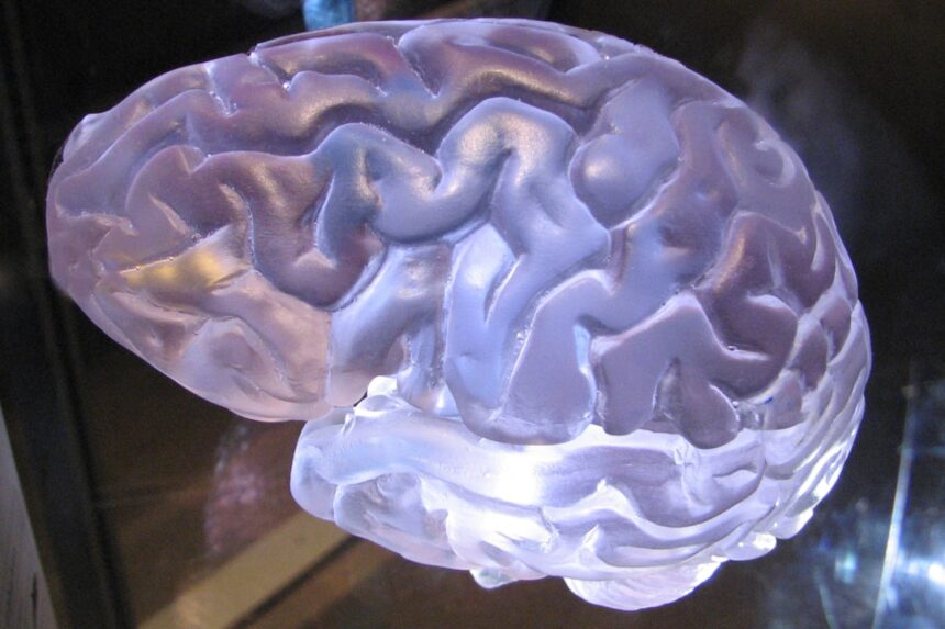
As you delve into the intricate world of the human brain, you quickly realize that it is one of the most complex structures known to science. Comprising approximately 86 billion neurons, each connected by synapses that can number in the thousands, the brain operates as a highly sophisticated network. This complexity is not merely a matter of quantity; it also involves the diverse functions these neurons perform, from regulating basic bodily functions to enabling higher cognitive processes such as reasoning, creativity, and emotional regulation.
The brain’s architecture is a marvel of evolution, designed to adapt and respond to an ever-changing environment. You may find it fascinating that the brain is not a static organ; it is dynamic and capable of remarkable plasticity. This means that your experiences, learning, and even injuries can reshape its structure and function over time.
Understanding this complexity is crucial for neuroscientists who seek to unravel the mysteries of how the brain works. By studying its various regions and their interconnections, researchers aim to gain insights into everything from memory formation to the development of neurological disorders. The quest to understand the brain is not just an academic pursuit; it has profound implications for health, education, and even artificial intelligence. Here is the sentence with the link:
You can watch a video about Cotard Delusion explained at
As you navigate through the landscape of brain mapping, you will discover a variety of techniques, each with its unique strengths and applications. These methods can be broadly categorized into structural imaging techniques, which provide detailed images of brain anatomy, and functional imaging techniques, which reveal how different areas of the brain activate during various tasks. Understanding these distinctions is crucial for appreciating how each technique contributes to our overall knowledge of brain function.
Structural imaging techniques include methods like magnetic resonance imaging (MRI) and computed tomography (CT) scans. These approaches allow you to visualize the physical structure of the brain, identifying abnormalities such as tumors or lesions. On the other hand, functional imaging techniques like functional MRI (fMRI) and positron emission tomography (PET) focus on measuring brain activity by detecting changes in blood flow or metabolic processes.
By combining these different types of imaging, researchers can gain a comprehensive understanding of both the anatomy and functionality of the brain.
Functional Magnetic Resonance Imaging (fMRI)
| Metrics | Value |
|---|---|
| Resolution | 1-3 mm |
| Temporal Resolution | 1-2 seconds |
| Spatial Coverage | Whole brain |
| Signal-to-Noise Ratio | High |
When you think about functional magnetic resonance imaging (fMRI), consider it one of the most groundbreaking advancements in neuroscience. This technique measures brain activity by detecting changes in blood flow associated with neural activity. When a specific area of your brain becomes more active, it requires more oxygen-rich blood, which fMRI can visualize in real-time.
This capability allows researchers to create dynamic maps of brain activity while you engage in various tasks or experiences. The applications of fMRI are vast and varied. In research settings, you might find it used to study everything from language processing to emotional responses.
For instance, by observing which areas of your brain light up when you hear words or see images, scientists can gain insights into how language is processed or how memories are formed. In clinical settings, fMRI can assist in pre-surgical planning for patients with epilepsy or tumors by identifying critical areas that must be preserved during surgery. The ability to visualize brain function in such detail has opened new avenues for understanding both normal cognitive processes and pathological conditions.
Diffusion Tensor Imaging (DTI)
As you explore further into brain mapping techniques, diffusion tensor imaging (DTI) stands out as a specialized form of MRI that focuses on the brain’s white matter tracts.
This diffusion is influenced by the orientation and integrity of axons—the long projections from neurons that connect different parts of the brain.
By analyzing this diffusion pattern, DTI can reveal important information about the connectivity between different brain regions. DTI has significant implications for understanding various neurological conditions. For example, if you consider traumatic brain injuries or neurodegenerative diseases like multiple sclerosis, DTI can help identify disruptions in white matter integrity that may correlate with cognitive deficits or other symptoms.
Researchers are increasingly using DTI to study developmental changes in the brain as well, providing insights into how connectivity evolves from childhood through adulthood. This technique not only enhances our understanding of normal brain development but also sheds light on how disruptions can lead to disorders.
Electroencephalography (EEG)

Electroencephalography (EEG) is another vital tool in your exploration of brain mapping techniques. This method involves placing electrodes on your scalp to measure electrical activity produced by neurons firing in your brain. EEG provides a direct measure of neural activity with excellent temporal resolution, allowing researchers to track changes in brain waves over milliseconds.
This capability makes EEG particularly valuable for studying processes that occur rapidly, such as sensory perception or cognitive tasks. One of the most compelling aspects of EEG is its application in clinical settings. For instance, if you were experiencing seizures or unexplained neurological symptoms, an EEG could help identify abnormal electrical patterns associated with epilepsy or other conditions.
Additionally, EEG is often used in sleep studies to analyze sleep stages and diagnose disorders like sleep apnea or insomnia. The ability to capture real-time electrical activity makes EEG an indispensable tool for both research and clinical practice.
Magnetoencephalography (MEG)
As you continue your journey through brain mapping technologies, magnetoencephalography (MEG) emerges as a powerful technique that complements EEG by measuring magnetic fields generated by neuronal activity. While EEG captures electrical signals from the scalp, MEG provides a more precise localization of brain activity due to its sensitivity to magnetic fields produced by synchronized neuronal firing. This allows researchers to pinpoint where in your brain specific activities are occurring with remarkable accuracy.
MEG has proven invaluable in both research and clinical contexts. In research settings, it is often used to study sensory processing and cognitive functions such as attention and memory. For example, if you were participating in an experiment designed to assess how your brain responds to visual stimuli, MEG could help identify which areas are activated during this process.
Clinically, MEG is increasingly being utilized for pre-surgical mapping in patients with epilepsy or tumors, helping surgeons avoid critical areas that could impact motor or language functions during surgery.
Positron Emission Tomography (PET)
Positron emission tomography (PET) represents another significant advancement in brain mapping technology that allows you to visualize metabolic processes within the brain. Unlike fMRI or EEG, which primarily measure blood flow or electrical activity, PET uses radioactive tracers injected into your bloodstream to detect metabolic activity in different regions of the brain. This technique provides insights into how various areas function under different conditions or during specific tasks.
PET scans have been instrumental in advancing our understanding of neurodegenerative diseases such as Alzheimer’s disease. By measuring glucose metabolism in the brain, PET can help identify early signs of cognitive decline before structural changes become apparent on MRI scans. Additionally, PET is often used in research settings to study neurotransmitter systems and their role in mental health disorders like depression or schizophrenia.
The ability to visualize metabolic activity adds another layer of understanding to how the brain operates and responds to various stimuli.
The Future of Brain Mapping Technology
As you look ahead into the future of brain mapping technology, it becomes clear that advancements are occurring at an unprecedented pace. Researchers are continually developing new techniques and refining existing ones to enhance our understanding of the human brain further. Innovations such as high-resolution imaging methods and machine learning algorithms are poised to revolutionize how we analyze and interpret complex data sets generated by various imaging modalities.
Moreover, there is growing interest in integrating multiple imaging techniques to create a more comprehensive picture of brain function and connectivity. For instance, combining fMRI with DTI could provide insights into both functional activation patterns and structural connectivity within the same individual. As technology continues to evolve, you can expect even more sophisticated tools that will allow researchers and clinicians to explore previously uncharted territories within the human mind.
Applications of Brain Mapping in Medicine and Psychology
The applications of brain mapping extend far beyond basic research; they have profound implications for medicine and psychology as well. In clinical settings, these techniques are increasingly being used for diagnostic purposes, treatment planning, and monitoring disease progression. For example, if you were diagnosed with a neurological disorder such as Parkinson’s disease or multiple sclerosis, advanced imaging could help tailor treatment strategies based on your unique brain structure and function.
In psychology, brain mapping has opened new avenues for understanding mental health disorders and their underlying neurobiological mechanisms. By identifying specific patterns of brain activity associated with conditions like anxiety or depression, researchers can develop targeted interventions that address these issues more effectively. Furthermore, neurofeedback—a technique that uses real-time data from EEG or fMRI—has emerged as a promising therapeutic approach for various psychological conditions by allowing individuals to learn how to regulate their own brain activity.
Ethical Considerations in Brain Mapping Research
As you engage with the exciting world of brain mapping technology, it is essential to consider the ethical implications that accompany these advancements. The ability to visualize and manipulate brain function raises important questions about privacy, consent, and potential misuse of information. For instance, if researchers can identify specific neural correlates associated with thoughts or behaviors, what safeguards are in place to protect individuals from unauthorized access to their mental states?
Additionally, there are concerns about how findings from brain mapping studies might be interpreted or misused in societal contexts—such as legal settings where neuroimaging results could influence judgments about culpability or responsibility. As neuroscience continues to advance rapidly, it becomes increasingly important for researchers and policymakers alike to engage in discussions about ethical standards that ensure responsible use of these powerful technologies while promoting public trust in scientific research. In conclusion, your journey through the realm of brain mapping reveals not only its complexity but also its transformative potential across various fields—from medicine to psychology and beyond.
As technology continues to evolve and our understanding deepens, you stand at the forefront of a new era in neuroscience that promises exciting discoveries and profound implications for humanity’s understanding of itself.
In exploring the intricate relationship between brain mapping and self-perception, a related article that delves into the psychological implications of self-awareness can be found on Unplugged Psych. This resource provides valuable insights into how our understanding of the brain can influence our sense of identity and personal growth. For more information, you can read the article [here](https://www.unpluggedpsych.com/).
WATCH THIS! Cotard Delusion: When Your Brain Deletes Your Soul
FAQs
What is brain mapping the self?
Brain mapping the self is a process of using neuroimaging techniques to understand how the brain represents the concept of self. It involves studying the neural correlates of self-referential processing and self-awareness.
Why is brain mapping the self important?
Understanding how the brain represents the self is important for gaining insights into various aspects of human cognition, behavior, and mental health. It can also help in understanding disorders related to self-awareness, such as schizophrenia and autism.
What techniques are used in brain mapping the self?
Neuroimaging techniques such as functional magnetic resonance imaging (fMRI), electroencephalography (EEG), and magnetoencephalography (MEG) are commonly used to study brain mapping the self. These techniques allow researchers to observe brain activity while individuals engage in self-referential tasks.
What are the potential applications of brain mapping the self?
The findings from brain mapping the self can have applications in fields such as psychology, psychiatry, neurology, and artificial intelligence. It can also contribute to the development of interventions for mental health disorders and the understanding of consciousness.
What are some challenges in brain mapping the self?
Challenges in brain mapping the self include the complexity of the concept of self, variability in individual self-representation, and the limitations of current neuroimaging techniques in capturing the intricacies of self-referential processing in the brain.